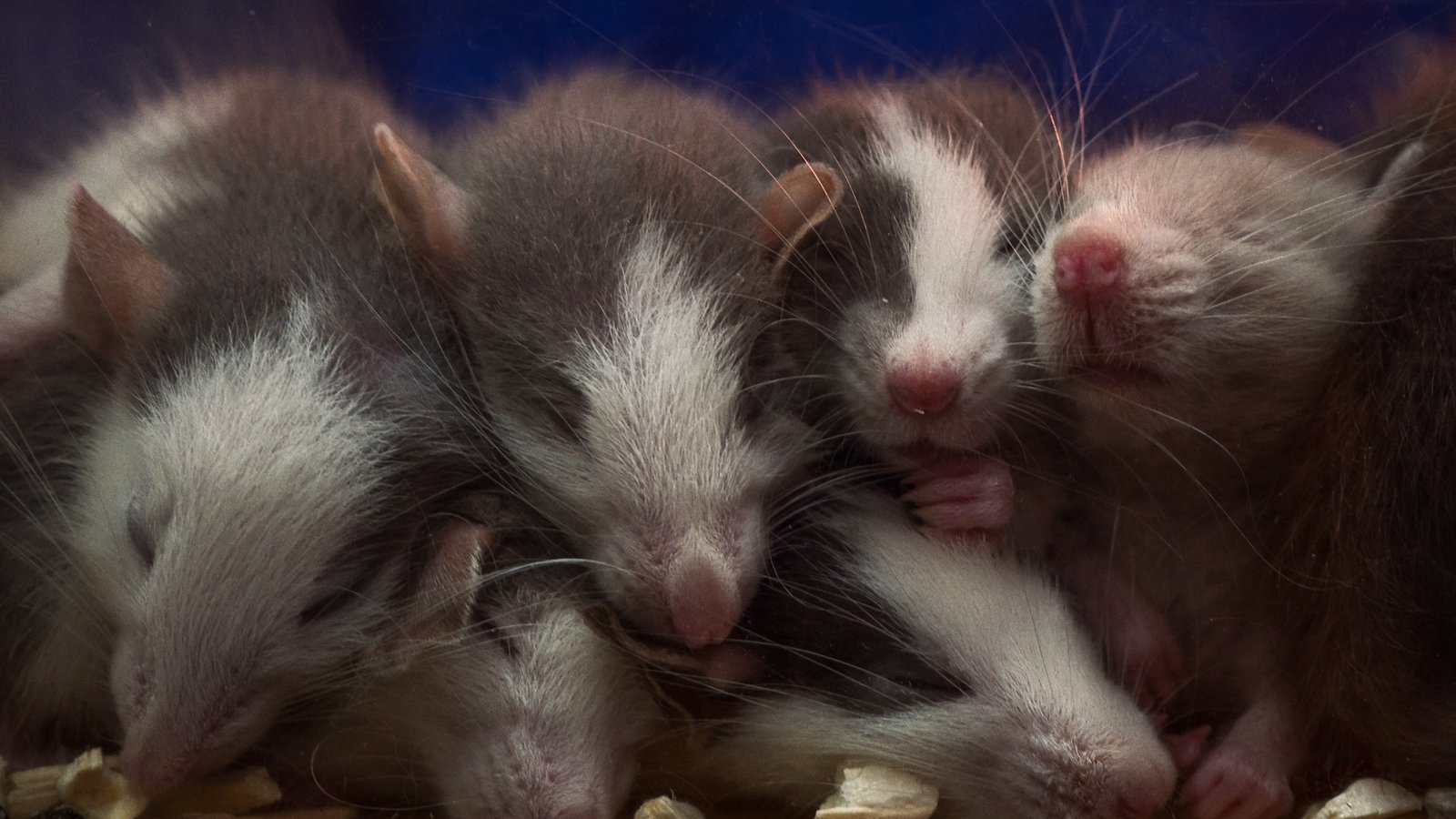

Iron deficiency during pregnancy can cause a male mouse embryo to develop female features, a new study reveals.
The low iron disrupts the activation of a key gene that spurs the development of male sex organs. This causes embryos with XY chromosomes — the most common combination seen in males — to develop female sex organs instead.
“This is a completely new and totally unexpected finding,” study co-author Peter Koopman, a professor emeritus of developmental biology at the University of Queensland in Australia, told Live Science. “It’s never been shown before that iron can flip such an important developmental switch.”
Earlier research established that the SRY gene on the Y chromosome is the “master switch” for turning on the development of male organs in mammals. An enzyme called JMJD1A plays an important role in flipping this master switch, and it requires iron to function properly. However, the connection between iron levels and sex determination was not fully understood.
Now, in a study published June 4 in the journal Nature, researchers report that iron is essential for the development of testes in XY mice. The results show that maternal iron deficiency disrupts the activity of JMJD1A, which lowers SRY expression and drives the development of ovaries in XY mouse embryos.
However, it’s too early to say whether this finding in mice might translate to human pregnancy and sex development, Tony Gamble, an associate professor of biological sciences at Marquette University in Milwaukee who wasn’t involved in the study, told Live Science.
In the study, the researchers used pharmaceutical treatments and low-iron diets to manipulate the iron levels in pregnant mice. When the pregnant mice experienced iron deficiency, this caused six out of 39 total XY embryos to develop ovaries instead of testes. Investigating further, they found that genetics appear to be a factor in which embryos are sensitive to this effect.
To confirm this mechanism, the team also grew embryonic gonads — structures that develop into testes or ovaries in the womb — in lab dishes so they could directly observe the impact of iron depletion. These lab analyses showed that reducing the iron in cells to 40% of normal levels led to a large increase in histones on the SRY gene. Histones are proteins that bind DNA and help control which genes are switched on, and this effect almost completely blocked the SRY gene’s expression.
Normally, the JMJD1A enzyme rids the SRY gene of histones, allowing it to turn on. The researchers hypothesize that when iron levels drop, the enzyme’s activity is compromised, so suppressive histones build up on the SRY gene.
These results suggest that “some important developmental traits that were previously thought to be purely genetically controlled can also be seriously impacted by nutrition and metabolic factors,” Koopman said. And “if iron can have such an impact on sex development, then maybe other organ systems may also critically depend on iron or other dietary factors in a similar way,” he added.
Because the research was conducted solely in mice, the question of whether iron may have similar effects in humans is still open. Although sex determination follows a broadly similar blueprint across mammals, there are some important differences between mice and humans, Gamble said.
For example, while both species rely on the same genes to drive the development of testes, the consequences of mutations in these genes differ between the two species. Their similarities to humans make mice important models for studying development and disease, Gamble said, “but the differences urge caution in simply assuming processes are acting identically across both species.”
Testing the new finding in humans won’t be easy, since many of the experiments possible in mice can’t ethically be done in humans, Koopman said. “So, the way forward will have to involve doing biochemical, cell culture and gene expression experiments to build a body of indirect evidence that what holds true in mice is also the case in humans,” he said.